Scatter segmentation in dynamic SPECT images using principal component analysis


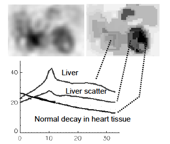
Dynamic single photon emission computed tomography (dSPECT) provides time-varying spatial information about changes of tracer distribution in the body from data acquired using a standard (single slow rotation) protocol. Variations of tracer distribution observed in the images might be due to physiological processes in the body, but may also stem from reconstruction artefacts. These two possibilities are not easily separated because of the highly underdetermined nature of the dynamic reconstruction problem. Since it is expected that temporal changes in tracer distribution may carry important diagnostic information, the analysis of dynamic SPECT images should consider and use this additional information. In this paper we present a segmentation scheme for aggregating voxels with similar time activity curves (TACs). Voxel aggregates are created through region merging based on a similarity criterion on a reduced set of features, which is derived after transformation into eigenspace. Region merging was carried out on dSPECT images from simulated and patient myocardial perfusion studies using various stopping criteria and ranges of accumulated variances in eigenspace. Results indicate that segmentation clearly separates heart and liver tissues from the background. The segmentation quality did not change significantly if more than 99% of the variance was incorporated into the feature vector. The heart behaviour followed an expected exponential decay curve while some variation of time behaviour in liver was observed. Scatter artefacts from photons originating from liver could be identified in long as well as in short studies.
 Top
Top
- Toennies, Klaus D.
- Celler, Anna
- Blinder, Stephan
- Möller, Torsten
- Harrop, Ronald
Top
Category |
Paper in Conference Proceedings or in Workshop Proceedings (Full Paper in Proceedings) |
Event Title |
SPIE International Symposium on Medical Imaging |
Divisions |
Visualization and Data Analysis |
Subjects |
Computergraphik |
Event Location |
San Diego, California |
Event Type |
Conference |
Event Dates |
Feb 15-20 |
Date |
February 2003 |
Export |
Top
