Segmentation-based regularization of dynamic SPECT reconstruction


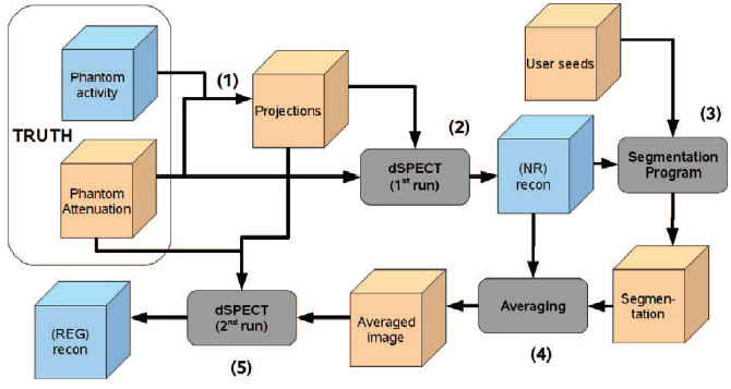
Dynamic SPECT reconstruction using a single slow camera rotation is a highly underdetermined problem, which requires the use of regularization techniques to obtain useful results. The dSPECT algorithm (Farncombe et al. 1999) provides temporal but not spatial regularization, resulting in poor contrast and low activity levels in organs of interest, due mostly to blurring. In this paper we incorporate a user-assisted segmentation algorithm (Saad et al. 2008) into the reconstruction process to improve the results. Following an initial reconstruction using the existing dSPECT technique, a user places seeds in the image to indicate regions of interest (ROIs). A random-walk based automatic segmentation algorithm then assigns every voxel in the image to one of the ROIs, based on its proximity to the seeds as well as the similarity between time activity curves (TACs). The user is then able to visualize the segmentation and improve it if necessary. Average TACs are extracted from each ROI and assigned to every voxel in the ROI, giving an image with a spatially uniform TAC in each ROI. This image is then used as initial input to a second run of dSPECT, in order to adjust the dynamic image to better fit the projection data. We test this approach with a digital phantom simulating the kinetics of Tc99m-DTPA in the renal system, including healthy and unhealthy behaviour. Summed TACs for each kidney and the bladder were calculated for the spatially regularized and non-regularized reconstructions, and compared to the true values. The TACs for the two kidneys were noticeably improved in every case, while TACs for the smaller bladder region were unchanged. Furthermore, in two cases where the segmentation was intentionally done incorrectly, the spatially regularized reconstructions were still as good as the non-regularized ones. In general, the segmentation-based regularization improves TAC quality within ROIs, as well as image contrast.
 Top
Top
- Humphries, T.
- Saad, A.
- Celler, A.
- Hamarneh, G.
- Möller, Torsten
- Trummer, M.R.
Top
Category |
Paper in Conference Proceedings or in Workshop Proceedings (Poster in Proceedings) |
Event Title |
IEEE Nuclear Science Symposium and Medical Imaging Conference 2009 |
Divisions |
Visualization and Data Analysis |
Event Location |
Orlando, FL, USA |
Event Type |
Conference |
Event Dates |
Oct. 24 2009-Nov. 1 2009 |
Page Range |
pp. 2849-2852 |
Date |
2009 |
Official URL |
http://dx.doi.org/10.1109/NSSMIC.2009.5401638 |
Export |
Top
